8U3Z

Model of TTLL6 bound to microtubule from composite map


Experimental Data Snapshot

  • Method: ELECTRON MICROSCOPY
  • Resolution: 3.60 Å
  • Aggregation State: FILAMENT 
  • Reconstruction Method: SINGLE PARTICLE 

wwPDB Validation   3D Report Full Report


This is version 1.0 of the entry. See complete history


Literature

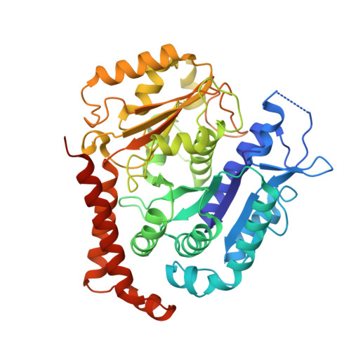
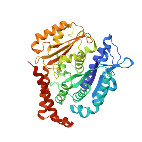
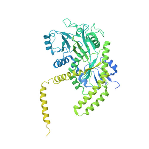
Structural basis for alpha-tubulin-specific and modification state-dependent glutamylation.

Mahalingan, K.K.Grotjahn, D.A.Li, Y.Lander, G.C.Zehr, E.A.Roll-Mecak, A.

(2024) Nat Chem Biol 

  • DOI: https://doi.org/10.1038/s41589-024-01599-0
  • Primary Citation of Related Structures:  
    8T42, 8U3Z

  • PubMed Abstract: 

    Microtubules have spatiotemporally complex posttranslational modification patterns. Tubulin tyrosine ligase-like (TTLL) enzymes introduce the most prevalent modifications on α-tubulin and β-tubulin. How TTLLs specialize for specific substrate recognition and ultimately modification-pattern generation is largely unknown. TTLL6, a glutamylase implicated in ciliopathies, preferentially modifies tubulin α-tails in microtubules. Cryo-electron microscopy, kinetic analysis and single-molecule biochemistry reveal an unprecedented quadrivalent recognition that ensures simultaneous readout of microtubule geometry and posttranslational modification status. By binding to a β-tubulin subunit, TTLL6 modifies the α-tail of the longitudinally adjacent tubulin dimer. Spanning two tubulin dimers along and across protofilaments (PFs) ensures fidelity of recognition of both the α-tail and the microtubule. Moreover, TTLL6 reads out and is stimulated by glutamylation of the β-tail of the laterally adjacent tubulin dimer, mediating crosstalk between α-tail and β-tail. This positive feedback loop can generate localized microtubule glutamylation patterns. Our work uncovers general principles that generate tubulin chemical and topographic complexity.


  • Organizational Affiliation

    Cell Biology and Biophysics Unit, Porter Neuroscience Research Center, National Institute of Neurological Disorders and Stroke, Bethesda, MD, USA.


Macromolecules
Find similar proteins by:  (by identity cutoff)  |  3D Structure
Entity ID: 1
MoleculeChains Sequence LengthOrganismDetailsImage
Tubulin alpha-1B chainA,
D [auth F]
451Homo sapiensMutation(s): 0 
UniProt & NIH Common Fund Data Resources
Find proteins for P68363 (Homo sapiens)
Explore P68363 
Go to UniProtKB:  P68363
PHAROS:  P68363
GTEx:  ENSG00000123416 
Entity Groups  
Sequence Clusters30% Identity50% Identity70% Identity90% Identity95% Identity100% Identity
UniProt GroupP68363
Sequence Annotations
Expand
  • Reference Sequence
Find similar proteins by:  (by identity cutoff)  |  3D Structure
Entity ID: 2
MoleculeChains Sequence LengthOrganismDetailsImage
Tubulin beta chainB,
C [auth E]
444Homo sapiensMutation(s): 0 
UniProt & NIH Common Fund Data Resources
Find proteins for P07437 (Homo sapiens)
Explore P07437 
Go to UniProtKB:  P07437
PHAROS:  P07437
GTEx:  ENSG00000196230 
Entity Groups  
Sequence Clusters30% Identity50% Identity70% Identity90% Identity95% Identity100% Identity
UniProt GroupP07437
Sequence Annotations
Expand
  • Reference Sequence
Find similar proteins by:  (by identity cutoff)  |  3D Structure
Entity ID: 3
MoleculeChains Sequence LengthOrganismDetailsImage
Tubulin polyglutamylase TTLL6E [auth N]822Mus musculusMutation(s): 0 
Gene Names: Ttll6
EC: 6.3.2
UniProt & NIH Common Fund Data Resources
Find proteins for A4Q9E8 (Mus musculus)
Explore A4Q9E8 
Go to UniProtKB:  A4Q9E8
IMPC:  MGI:2683461
Entity Groups  
Sequence Clusters30% Identity50% Identity70% Identity90% Identity95% Identity100% Identity
UniProt GroupA4Q9E8
Sequence Annotations
Expand
  • Reference Sequence
Small Molecules
Ligands 4 Unique
IDChains Name / Formula / InChI Key2D Diagram3D Interactions
GTP
Query on GTP

Download Ideal Coordinates CCD File 
F [auth A],
L [auth F]
GUANOSINE-5'-TRIPHOSPHATE
C10 H16 N5 O14 P3
XKMLYUALXHKNFT-UUOKFMHZSA-N
G2P (Subject of Investigation/LOI)
Query on G2P

Download Ideal Coordinates CCD File 
H [auth B],
J [auth E]
PHOSPHOMETHYLPHOSPHONIC ACID GUANYLATE ESTER
C11 H18 N5 O13 P3
GXTIEXDFEKYVGY-KQYNXXCUSA-N
ATP
Query on ATP

Download Ideal Coordinates CCD File 
N
ADENOSINE-5'-TRIPHOSPHATE
C10 H16 N5 O13 P3
ZKHQWZAMYRWXGA-KQYNXXCUSA-N
MG
Query on MG

Download Ideal Coordinates CCD File 
G [auth A],
I [auth B],
K [auth E],
M [auth F],
O [auth N]
MAGNESIUM ION
Mg
JLVVSXFLKOJNIY-UHFFFAOYSA-N
Experimental Data & Validation

Experimental Data

  • Method: ELECTRON MICROSCOPY
  • Resolution: 3.60 Å
  • Aggregation State: FILAMENT 
  • Reconstruction Method: SINGLE PARTICLE 
EM Software:
TaskSoftware PackageVersion
MODEL REFINEMENTPHENIX
RECONSTRUCTIONRELION3.0

Structure Validation

View Full Validation Report


Entry History & Funding Information

Deposition Data


Funding OrganizationLocationGrant Number
National Institutes of Health/National Institute of Neurological Disorders and Stroke (NIH/NINDS)United StatesZO1 NS003163

Revision History  (Full details and data files)

  • Version 1.0: 2024-05-29
    Type: Initial release